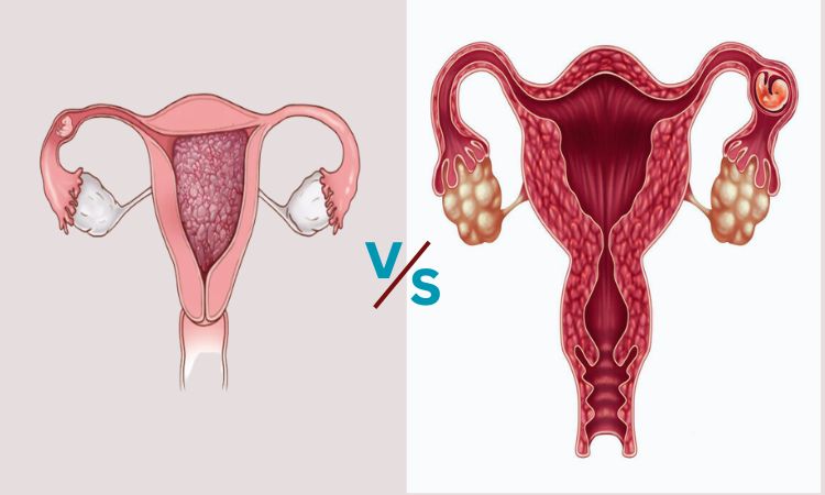
Hydatidiform Mole vs. Ectopic Pregnancy
Hydatidiform Mole: Hydatidiform moles are rare pregnancy-related disorders characterized by abnormal tissue growth within the uterus. They can be divided into full and partial forms depending on genetic abnormalities, with symptoms including vaginal bleeding, an enlarged uterus, and no fetal heart sounds.
Diagnosis usually includes ultrasound imaging followed by monitoring of HCG levels, then removal of abnormal tissues through dilation and curettage with subsequent monitoring to avoid potential malignancy risks.
Ectopic Pregnancy: Ectopic pregnancy occurs when fertilized eggs implant outside of the uterus, most frequently in the fallopian tubes. It poses serious health risks, including abdominal pain, vaginal bleeding, and positive pregnancy tests. Transvaginal ultrasound and hCG level monitoring help detect the condition.
Stable cases may be treated with medication while unstable cases require surgical intervention to avoid rupture complications or rupture complications that require prompt diagnosis and management to avoid potentially life-threatening situations.
What is Hydatidiform Mole?
Hydatidiform moles, commonly referred to as molar pregnancy are rare yet potentially serious pregnancy-related conditions that result in abnormal placental tissue development that creates grape-like clusters instead of healthy fetuses. Complete and partial hydatidiform moles exist. In a complete mole, there is no normal fetus present and its placental tissue develops from genetic material from duplicated sperm cells.
Partial moles arise when genetic material from both parents is combined, yet does not result in a viable fetus. Symptoms may include vaginal bleeding, an enlarged uterus, and elevated levels of the hormone hCG.
Diagnosis is typically accomplished via ultrasound monitoring Treatment typically entails the removal of abnormal tissues followed by close monitoring for any complications or malignancy risks.
what is Ectopic Pregnancy?
The condition of ectopic pregnancy can be a grave and life-threatening problem where fertilized eggs are implanted out of the uterus, and begin to grow outside the uterus typically inside the fallopian tubes. An ectopic pregnancy differs from its counterpart in that the fertilized egg fails to implant correctly into the uterus, rather, in an ectopic pregnancy it cannot survive and develop properly within its fallopian tube or elsewhere abnormal locations.
This condition poses serious health risks to mothers due to its potential for fallopian tube rupture and internal bleeding, often manifesting itself through abdominal pain, vaginal bleeding, and positive pregnancy tests. Common symptoms include abdominal and vaginal pain as well as positive pregnancy tests.
Diagnosis of an ectopic pregnancy can often be determined through transvaginal ultrasound and monitoring of hCG levels, with treatment options including medications or surgical removal depending on its stability. Rapid diagnosis and appropriate management are crucial in order to reduce complications and safeguard maternal well-being.
Symptoms and Causes
Symptoms of Hydatidiform Mole
- Vaginal Bleeding: It is usually unpredictable and can be more than usual.
- An enlarged Uterus: Itis when the uterus becomes bigger than what is expected at this phase of pregnancy.
- Absence of Fetal Heart sounds: In the absence of Fetal Heart sounds, during the auscultation test, there is no sign of a heartbeat from an unborn fetus can be detected.
- Urgent nausea and vomiting: Hyperemesis Gravidarum, extreme nausea, and vomiting can be an indication.
- The hCG levels: It elevated patients are higher than normal levels of the human chorionic gonadotropin (hCG) hormone. It is commonly observed in pregnancy tests.
Symptoms of Ectopic Pregnancy
- Abdominal Pain: Sharp and extreme pain in one part of the abdominal, or pelvis. Usually, it gets worse in time.
- Vaginal bleeding: Light to large spotting or bleeding typically darker than normal menstrual blood.
- Shoulder pain: Rarely, painful shoulder pain can be caused by bleeding that is affecting the diaphragm.
- Urination Pain: The pain of urination and bowel movements In the case of discomfort when urinating, or the bowel movement can occur.
- Frenzy or fainting: due to internal bleeding as well as low blood pressure.
- Positive Test for Pregnancy: The pregnancy test has been positive, however, the signs suggest that the location of your pregnancy may be abnormal.
What causes Hydatidiform Mole
Hydatidiform moles usually result from a problem with fertilization which is when the genetic material of the egg and sperm are combined incorrectly. When a mole is completely hydatidiform it is the case that the egg does not contain genetic material and the genetic material of sperm duplicates and causes the growth of abnormally large placental tissues, with no viable fetus.
In a partial hydatidiform mole eggs are fertilized by two sperm, or a normal sperm, and an irregular sperm. This causes an irregular development of the fetus and the abnormal growth of the placenta.
What causes Ectopic Pregnancy
Ectopic pregnancies result when fertilized eggs are implanted outside of the uterus, typically in the fallopian tube.
Many factors can cause the abnormality of this implantation:
- Injured Fallopian Tubes: The scar tissues from surgeries, infections, or other conditions such as endometriosis may hinder eggs’ passage through the fallopian tubes which can lead to an ectopic birth.
- Hormonal Imbalance: Discords in the levels of hormones can impact the flow of fertilized egg inside the fallopian tube, which can cause it to go into the wrong place.
- Infections and inflammation: Pelvic inflammatory disease (PID) or sexually transmitted infections may cause inflammation and scarring within the fallopian tubes, causing disruption to the flow of eggs, as well as increasing the likelihood of having an ectopic pregnancy.
- Previous Ectopic Pregnancy: A woman who has experienced an ectopic pregnancy prior to this has a greater chance of having a second.
- Reproductive Health Disorders: Issues such as adenomyosis and uterine fibroids could hinder eggs’ transfer to the uterus and raise the risk of an ectopic pregnancy.
Comparison chart between Hydatidiform Mole and Ectopic Pregnancy
Here’s a comparison chart outlining the key differences between Hydatidiform Mole and Ectopic Pregnancy:
| Aspect | Hydatidiform Mole | Ectopic Pregnancy |
|---|---|---|
| Definition | Abnormal growth of placental tissue | Implantation of embryo outside |
| within the uterus | the uterus | |
| Types | Complete and Partial Mole | Various sites, commonly in |
| fallopian tubes | ||
| Presence of Fetus | Absent (complete mole), Abnormal | Abnormal development outside |
| fetal tissue (partial mole) | uterus | |
| Clinical Symptoms | Vaginal bleeding, enlarged uterus, | Abdominal pain, vaginal bleeding, |
| absence of fetal heart sounds | positive pregnancy test | |
| Diagnosis Methods | Ultrasound imaging, hCG level | Transvaginal ultrasound, hCG |
| monitoring | level monitoring | |
| Management | Removal of abnormal tissue via D&C, | Medication (methotrexate) for |
| close monitoring for malignancy | stable cases; surgery for unstable | |
| Potential Complications | Risk of developing gestational | Risk of fallopian tube rupture, |
| and Risks | trophoblastic neoplasia (cancer), | internal bleeding, potential |
| potential for malignancy | complications | |
| Long-Term Effects | Potential for recurrent disease | Increased risk of future |
| ectopic pregnancies | ||
| Importance of Early | Proper management to prevent | Early diagnosis and management to |
| Diagnosis and | malignancy, psychological impact on | prevent complications and ensure |
| Management | patient | maternal well-being |
Diagnosis and treatment
Diagnostic and Treatment Strategies for Hydatidiform Mole
Diagnosis:
- Ultrasonography: Uterine ultrasound images may reveal grape-like clusters associated with hydatidiform moles.
- Monitoring Human Chorionic Gonadotropin (hCG) Levels: Accurate diagnosis is dependent upon accurately measuring human chorionic gonadotropin (hCG) hormone levels, with higher-than-normal levels not supporting an identifiable pregnancy providing additional proof.
Treatment:
- Tissue Analysis: Following removal using dilation and curettage (D&C), the removed tissue is examined to confirm the diagnosis and assess potential malignancy.
- Treatment Options: Once Malignancy has been diagnosed, any necessary interventions such as surgery must take place immediately to remove and treat the affected areas.
- Dilation and Curettage (D&C): Dilation and Curettage is the primary treatment, which involves surgically extracting any abnormal placental tissue from within the uterus through dilation and curettage (D&C).
- Follow-Up Care: Following any procedure, regular monitoring of hCG levels must take place in order to confirm that all abnormal tissue has been eliminated and detect any potential signs of malignancy.
- Emotional Support: Psychological support can be crucial when living with this condition which can have severe psychological ramifications on patients. Diagnosis and
Diagnostic and Treatment Strategies for Ectopic Pregnancy
Diagnosis:
- Transvaginal Ultrasound: Imaging the pelvic area helps visualize where an embryo has implanted itself outside the uterus.
- HCG Levels: It’s important to monitor HCG levels closely – any slower-than-usual rise may indicate an ectopic pregnancy.
- Culdocentesis: When indicated, a needle may be inserted into the area behind the uterus (cul-de-sac) to detect fluid and identify internal bleeding.
Treatment:
- Medication: Medication may help. In stable cases where an embryo has caused no significant harm, methotrexate (a medication that prevents cell division) may be administered to dissolve an ectopic pregnancy.
- Surgery: When embryo damage has occurred, surgical intervention is sometimes necessary to ensure a safe gestation process. Laparoscopic surgery is often utilized for this procedure in order to safely remove ectopic tissues or damaged fallopian tubes.
- Follow-Up: Following treatment, closely monitoring hCG levels and recovery is important to achieving full resolution and avoiding complications.
Summary
Hydatidiform Mole is an abnormal form of placental development in the uterus and Ectopic Pregnancy occurs the time when fertilized eggs implant outside of the uterus, usually within the fallopian tube.
Hydatidiform Mole does not have a viable fetus and Ectopic Pregnancy is a risk because of the abnormality in the implant. Both require timely diagnostics and customized management to protect maternal health.