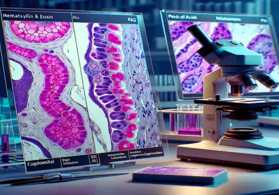
H&E and PAS Stain are similar to paints that are made for cells. H&E transforms cells into vibrant which aids in the diagnosis of ailments. PAS can identify sugars within cells, which is useful in treating liver diseases and infections.
H&E is the abbreviation to mean Hematoxylin as well as Eosin stain. It’s a unique coloring technique employed in medicine and science to aid in observing tissues and cells better under microscopes.
Hematoxylin causes the cell’s nuclei (the cell’s control center) change to blue. Eosin causes other components of cells, including the cytoplasm change color, either red or pink. H&E staining aids researchers and doctors analyze tissues and diagnose illnesses.
PAS is the acronym as a Periodic Acid-Schiff stain. The stain is utilized in a similar manner as H&E however it is particularly effective in highlighting sugars, such as glycogen, found in tissues. PAS staining causes sugars in cells change to a dark magenta hue. It’s useful for identifying glycogen stored in liver cells, as well as identifying diseases caused by sugar-coated bacteria.
What is H&E Stain?
Haematoxylin and eosin stain or H&E, which stands in short for Hematoxylin and Eosin is a vital staining technique that is used in medicine and science to improve the visibility of tissues and cells under microscopes. Consider it an specialized paint that aids scientists and doctors to know what’s going on inside our bodies’ tiny structures.
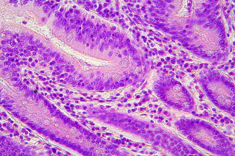
Hematoxylin is similar to an blue paintbrush. It emphasizes the nuclei of cells, which are the cells’ control centers. In contrast, Eosin acts like a paint that is red or pink and colors the other cell’s parts. This coloring makes it simpler for doctors and scientists to study tissues, detect the different cell structures and to diagnose illnesses.
H&E staining is an essential tool in histopathology that allows the identification of abnormalities and aiding in understanding different diseases. With H&E medical professionals can gain a greater understanding of the causes of problems in our bodies on the microscopic level, aiding in providing the best health care and treatment.
What is PAS Stain?
PAS, also known as the Periodic Acid-Schiff stain, can be an additional useful tool in the field of science and medicine. Think of it as a unique coloring technique, much like painting, but specifically for tissues and cells under microscope.
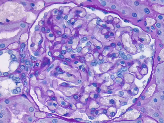
What distinguishes PAS distinct is its ability at the detection of sugars, like glycogen in cells. If you make use of PAS, these sugars transform into dark magenta in color and make them stick out.
PAS staining can be particularly helpful to identify stored sugars in cells, particularly in organs like the liver. It’s also helpful in finding certain illnesses caused by fungi or bacteria with sugary coatings. Simply put, PAS helps scientists and doctors to detect sugar-related elements in the microcosm of our body.
This method plays an important role in identifying various medical ailments and figuring out the way our bodies function on a micro-level. It’s similar to using a paintbrush to uncover subtle clues to our health.
Key Difference Between H&E and PAS Stain
Here’s a more detailed comparison chart with comprehensive information for each aspect of H&E (Hematoxylin and Eosin) staining and PAS (Periodic Acid-Schiff) staining:
| Aspect | H&E Staining | PAS Staining |
|---|---|---|
| Purpose | Used for general tissue and cell analysis in histopathology and research. | Primarily employed for detecting and visualizing carbohydrates, including glycogen and mucins, in tissues. |
| Components Stained | – Nuclei appear blue. | – Carbohydrates and mucins in tissues appear magenta. |
| Staining Mechanism | Hematoxylin binds to DNA and some proteins in the nuclei, while eosin stains cytoplasmic and extracellular components. | Periodic acid oxidizes carbohydrates in tissues, followed by reaction with Schiff’s reagent to produce the magenta color. |
| Color Appearance | – Nuclei: Blue. | – Carbohydrates and mucins: Magenta. |
| Routine Diagnostic Tool | Widely used in routine histopathology and research to provide a general overview of tissue architecture and pathology. | Typically not used as a routine diagnostic tool; PAS staining is reserved for specific diagnostic or research purposes related to carbohydrate detection. |
| Glycogen Detection | Not effective for glycogen detection. | Highly effective for detecting glycogen within tissues. |
| Common Use | Commonly employed for general tissue assessment, identifying abnormalities, and diagnosing diseases in clinical settings. | Primarily used in specialized diagnostics and research when detecting carbohydrates (e.g., glycogen storage diseases, mucopolysaccharidoses) or specific infectious agents is essential. |
| Versatility | Versatile and applicable to a wide range of tissue types and specimens. | Limited to carbohydrate-rich structures, making it less versatile for routine histopathology. |
| Application Challenges | Generally straightforward, with well-established protocols. | Involves a more complex procedure due to periodic acid treatment, necessitating precise timing and handling. |
| Clinical Significance | Provides valuable information for disease diagnosis and tissue assessment in clinical settings. | Critical for the diagnosis of glycogen storage diseases, certain infections, and diseases characterized by carbohydrate accumulation. |
| Counterstaining (Optional) | Hematoxylin is often used for counterstaining nuclei in H&E staining. | Hematoxylin may be used optionally for counterstaining nuclei in PAS staining. |
| Examination Under Microscope | Allows visualization of cellular details and tissue architecture. | Focuses on detecting carbohydrates and carbohydrate-rich structures under the microscope. |
| Safety Considerations | Standard laboratory safety precautions are required for handling staining chemicals. | Standard laboratory safety precautions are necessary when handling staining chemicals. |
Method of application of H&E and PAS Stain
H&E Stain Application:
- Preparing Tissue: Find a specimen of tissue then cut thin sections and place these on slides.
- Desparaffinization: Get rid of paraffin using xylene, and then rehydrate the pieces with alcohol.
- Hematoxylin: Stain nuclei blue using hematoxylin. wash.
- Eosin: Colorize cytoplasm red or pink using eosin. Rinse.
- Dehydrate and mount: Dehydrate gradually, then clear using xylene, then mount using the help of a coverslip.
- Review: Observe stained tissue sections under the microscope.
PAS Stain Application:
- Prepare Tissue: The same as H&E staining.
- Deparaffinization and Rehydration: Same as H&E.
- Periodic Acid: Utilize periodic acid in the treatment to reduce sugars.
- Schiff’s Reagent: Apply Schiff’s Reagent, incubate for 15-30 minutes to achieve a magenta-colored color.
- Sulfurous Acid: Cleanse the area with sulfurous acid in order to stop the process.
- Optional Counterstain: Use hematoxylin for nuclei (optional).
- Clear, Dehydrate, as well as Mount: Similar to H&E staining.
- Review: Observe stained tissue sections under a microscope in order to find PAS-positive or sugars.
Advantage and disadvantages
Advantages of H&E Staining
- Cellular Details: H&E staining provides an excellent visualisation of the cellular structure. It reveals cells’ nuclei (in blue) and the cytoplasm (in red or pink) which makes it much easier to analyze tissues and identify diseases.
- Multi-purpose: H&E staining is extensively applicable to a variety of specimens and tissues, which makes it a popular color for everyday pathology.
- Diagnostic Routine Tool: This is the common staining method used in pathology. It assists pathologists to identify abnormal and normal tissues, aiding in the diagnosis of cancer, as well as giving valuable information to make the treatment decision.
- Histopathological Analysis: Staining with H&E allows for the evaluation of tissue structure cells, types of cells, and inflammatory responses, making it possible an extensive histopathological analysis.
Disadvantages of H&E Staining
- Limited specificity: H&E staining gives an overall view of tissue structure, but does not provide specificity for certain cell components or other substances. It is not able to reveal particular structures such as mucins and glycogen.
- Limited Use in Certain diagnoses: H&E staining may not be enough to diagnose certain illnesses or conditions in which the presence of carbohydrate or other chemicals is vital.
- Not a good choice for glycogen detection: H&E staining is not efficient in highlighting glycogen in cells, an important limitation in diagnosing glycogen storage disorders.
- Color variation: Staining intensity and colors could depend on a variety of factors, including staining protocols, fixation techniques and the types of tissues that could cause inconsistencies with regards to the interpretation.
Advantages of PAS Staining
- Sugar Detection: PAS staining has been specifically created to detect sugars and carbohydrates within tissues. It also identifies mucins, glycogen and other structures that are rich in carbohydrate.
- Identification of Storage Diseases: PAS staining is crucial for diagnosing diseases involving the abnormal storage of glycogen or mucin, such as glycogen storage diseases and mucopolysaccharidoses.
- Infectious Agents: PAS staining may help identify infectious agents, such as parasites, fungi and certain bacteria that have glycogen-rich capsules. It can aid in diagnosing infections.
- Specific Applications: The technology is helpful in situations in which sugars’ presence needs to be verified, which makes it useful for specific studies and diagnostics.
Disadvantages of PAS Staining
- The use of HTML0 is limited to general usage: PAS staining is specifically designed for carbohydrate-rich tissues and does not offer an exhaustive image of the morphology of tissues. It is not appropriate for routine histopathological tests.
- Complex procedure: PAS staining involves multiple steps, which include periodic acid treatment and several rinses, which makes it more complicated and time-consuming in comparison to H&E staining.
- Interference: The PAS staining process can be affected due to several aspects, such as the size of the tissue sections as well as the quality of the reagents used that can cause staining artifacts.
- Interpretation Problems: Interpretation of the results of PAS staining can be difficult, because the coloration may vary and the distinction between different types of carbohydrates could require expertise.
- limited information: Although PAS staining can be excellent for detecting carbohydrate however, it doesn’t provide any information about other cell structures or components that could be crucial in a comprehensive analysis of tissues.
What are the Similarities Between H&E and PAS Stain
- Microscopic Examination: Both staining methods are employed to examine microscopically the cells and tissues.
- Histopathological Utilization: They are valuable tools in histopathology and aid to diagnose illnesses and in the examination of the structures of tissues.
- Staining techniques: Both use specific chemical staining methods to increase the visibility of cell components.
- Mounting of slides: Following staining both H&E and PAS stained slides are generally mounted with coverslips that allow examination under the microscope.
- Coloration: Each stain uses different colors (e.g. blue, red for H&E or magenta for PAS) to draw attention to specific cell characteristics.
- Clinical Application: Both are used in clinical settings to provide vital diagnostic data for the patient’s treatment.
Summary
H&E (Hematoxylin and Eosin) and PAS (Periodic Acid-Schiff) staining are specific coloration methods that are employed in medical and scientific research. H&E emphasizes cell structures using blue and red colors, which aids in the diagnosis of diseases.
Contrarily the PAS stain is great in revealing sugars inside cells, and is particularly effective to detect stored sugars and certain infections. Both stains play an essential part in analyzing the cells and tissues on the microscopic level. They contribute to improved health and disease diagnosis.